Cervical disk problems
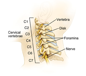
The spine has 3 natural curves. The cervical curve is in the neck. It forms the top part of the spine. This part is also known as the cervical spine. This sheet tells you more about the parts of the cervical spine and damaged disks. This is a common problem that can affect the cervical spine. Most people don't need surgery for this.
A healthy cervical spine
The spine is made up of these parts:
-
Vertebrae. These are bones stacked like building blocks that make up the spine. The neck contains the first 7 vertebrae of the spine.
-
Disks. These are small pads of tissue that lie between the vertebrae. They help cushion and protect the vertebrae and allow your spine to move.
-
The spinal cord. This nerve tissue runs through a large central opening (spinal canal) formed by the vertebrae.
-
Nerve root. These branch out from the spinal cord and carry signals to the body.
-
Foramina. These are smaller openings on each side of the vertebrae. Nerve roots and the nerves they form lead to your neck, shoulders, arms, and hands from the spinal cord through these openings.

Damaged disks in the cervical spine
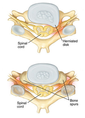
A common cervical spine problem is a damaged disk. A disk may be injured by a sudden movement. This can cause a disk to bulge or break open (herniate). Or a disk may wear out slowly over time (degenerate). A worn-out disk may become very flat. Then the vertebrae above and below it touch or slip back and forth. As disks wear out, abnormal bone growths (bone spurs) can form on the vertebrae. Bone spurs can also form in the foramina. This causes them to narrow (stenosis) and press on the nerve root.
If your healthcare provider thinks that you have a damaged disk, you may have tests to confirm the problem. You may have any of these:
Your healthcare provider will then work with you to plan treatment as needed.

Online Medical Reviewer:
Anne Fetterman RN BSN
Online Medical Reviewer:
Heather M Trevino BSN RNC
Online Medical Reviewer:
Mahammad Juber MD
Date Last Reviewed:
9/1/2024
© 2000-2025 The StayWell Company, LLC. All rights reserved. This information is not intended as a substitute for professional medical care. Always follow your healthcare professional's instructions.